Describe the Technique for Using Doppler Ultrasound
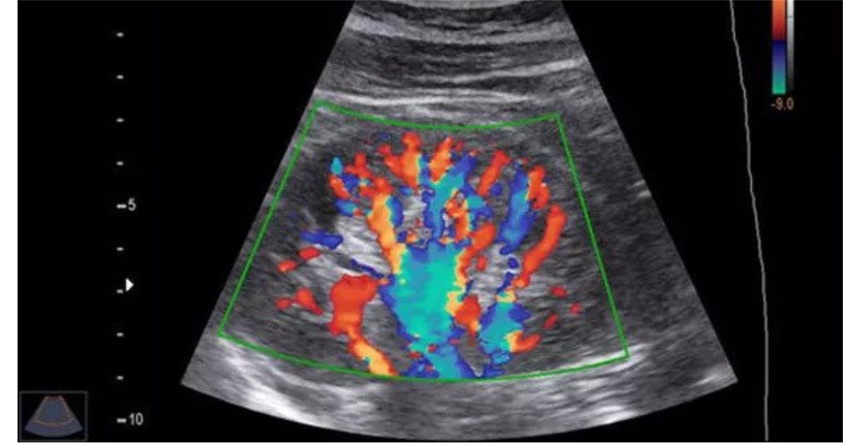
Vascular malformations and tumors comprise a wide heterogeneous spectrum of lesions that often represent a diagnostic and therapeutic challenge. Multiple parameters including gain and pulse repetition frequency need to be optimized to detect slow flow.
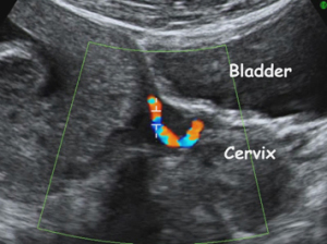
Doppler Ultrasound In Pregnancy Diagnostic Ultrasound
In addition to detect small-vessel lumen reductions the measurement of velocity changes is more sensitive than the measurement of flow.
. With this principle conventional Doppler techniques assess the velocity of blood flow by measuring high-frequency low-amplitude signals from small fast-moving blood cells. Color or power Doppler US requires proper technique to prevent erroneous interpretation. Spectral Doppler abnormalities can be used to identify obstruction in the vein segments central to the sample site.
The ultrasound Doppler technique is the only method that can feed rapid changes in cardiovascular conditions back in real time including the absolute mean and peak velocity of blood flow. Frequent use of an inaccurate nomenclature has led to considerable confusion. The presence of fetal movements and fetal heart rate acceleration is the most critical feature of the non-stress.
Patients Practice Policy Education. Randomized and quasi-randomized controlled trials of Doppler ultrasound for the investigation of utero-placental vessel waveforms in first and second trimesters compared with no Doppler ultrasound were included in this review. Imaging 84 2022 140-148 Jing Gao.
In some cases such as when color or power Doppler imaging demonstrates small foci of color rather than distinct vessels spectral Doppler imaging should. These researchers excluded studies where uterine vessels have been assessed together with fetal and umbilical vessels. A prenatal non-stress test functions in overall antepartum surveillance with ultrasound as a part or component of the biophysical profile.
Radiographic features of pneumonitis in. A preliminary observation Clin. Corrigendum to Ultrasound attenuation coefficient of the liver and spleen in adults.
Color Doppler may help identify smaller and pelvic veins in particular if augmentation is used. B-mode gray-scale imaging excels at defining the presence and extent of venous obstruction and color Doppler imaging facilitates differentiation of antegrade and retrograde blood flow. Since the treatment strategy depends on the type of vascular anomaly correct diagnosis and classification are crucial.
April 16 2022. Flexible electronic devices are soft. Color Doppler ultrasound can detect complete versus incomplete obstruction.
Doppler spectral waveform analysis is used to confirm venous blood flow direction and to demonstrate the presence or absence of phasic respiratory changes the amount of. It can be used to clarify otherwise technically difficult findings. Doppler echocardiography relies on detection of the shift in frequency of ultrasound signals reflected from moving objects.
In TDI the same Doppler principles are used to quantify the higher. Prenatal non-stress test popularly known as NST is a method used to test fetal wellbeing before the onset of labor.
4d Color Ultrasound Vikas Diagnostics
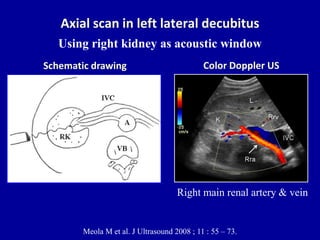
Doppler Ultrasound Of The Kidneys
Colour Doppler Test Cost Colour Doppler Ultrasound Price Near Me
No comments for "Describe the Technique for Using Doppler Ultrasound"
Post a Comment